What is Sonography?
Sonography is a diagnostic medical procedure that uses sound waves (ultrasound) to produce dynamic visual images of organs, tissues, or blood flow inside the body.
3D sonography involves creating a 3D image of the baby using sound waves. Unlike traditional 2D ultrasounds, which provide flat images, 3D sonography captures a series of images from different angles and then combines them to generate a 3D view.
4D sonography takes it a step further by adding the element of time, creating a moving 3D image or video of the fetus. This real-time view allows parents to see their baby's movements, gestures, and facial expressions, enhancing the emotional connection with the unborn child.
Why is Sonography needed?
The technology is comparable to radar and sonar, which are used by the military to find planes and ships. An ultrasound allows your doctor to see problems with organs, vessels, and tissues without needing to make an incision. Unlike other imaging techniques, ultrasound uses no radiation. For this reason, it’s the preferred method for viewing a developing fetus during pregnancy.
How is Sonography conducted?
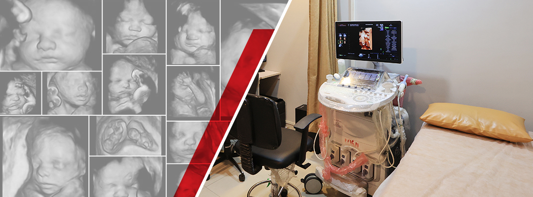
- A sonographer uses a specialized ultrasound machine equipped with a 3D/4D transducer.
- A water-based gel is applied to the mother's abdomen to ensure a clear acoustic window for the ultrasound waves.
- The transducer is moved over the abdomen to capture multiple images at various angles.
- The ultrasound machine processes these images to produce 3D or 4D images, which are then shown to the parents on a monitor.
At Vcare Sonography Centre in Kandivali, we incorporate 3D and 4D sonography into our services, empowering families to embark on their parenting journey with a profound understanding of their baby's growth and movements.