Imaging Tests During Pregnancy: What’s Safe and What’s Not
Introduction
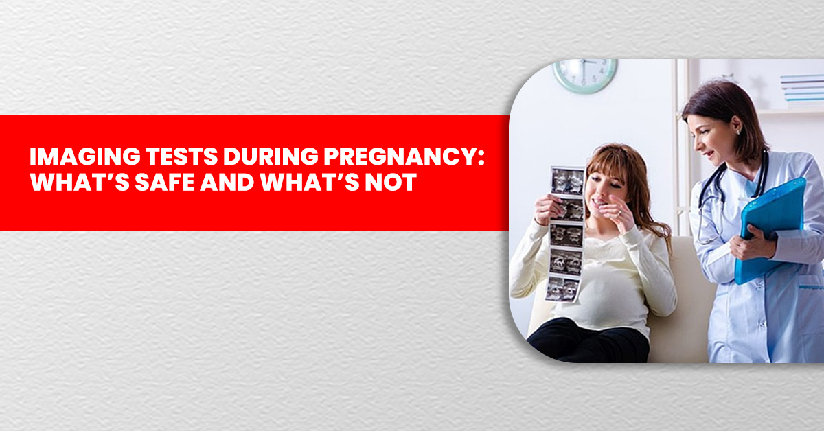
Motherhood is a precious journey filled with both excitement and concerns. Pregnancy is a transformative experience that brings significant changes to a woman’s body, making maternal and fetal health a top priority. Imaging tests play a crucial role in monitoring fetal development and detecting potential complications. However, expecting mothers and their families often worry about the safety of these tests. Understanding the risks, benefits, and precautions of imaging techniques such as ultrasounds, MRIs, X-rays, and CT scans at a CT scan centre in Kandivali can help make informed healthcare decisions.
Why Should One Do Imaging Tests During Pregnancy?
Monitoring Fetal Development
Imaging tests track the baby’s growth and development throughout pregnancy. Routine ultrasounds, including 3D and 4D sonography in Kandivali, allow healthcare providers to assess the baby's size, movements, and overall health. These tests also help detect congenital anomalies early, ensuring timely medical intervention.
Checking Fetal Position
As pregnancy progresses, determining the baby’s position becomes crucial, especially before delivery. Ultrasounds help identify whether the baby is in a breech or transverse position, allowing doctors to plan necessary medical or manual interventions for a safe delivery.
Assessing the Placenta and Amniotic Fluid
The placenta and amniotic fluid are essential for fetal development. Imaging tests help evaluate placental function and detect conditions like placenta previa or placental insufficiency. Doctors also assess amniotic fluid levels to prevent complications such as oligohydramnios (low fluid) or polyhydramnios (excess fluid).
Diagnosing High-Risk Pregnancy Complications
Women with high-risk pregnancies, including those with gestational diabetes, preeclampsia, or multiple pregnancies, require frequent imaging tests. Advanced imaging techniques help diagnose maternal and fetal conditions without exposing the baby to unnecessary risks.
Diagnosing Maternal Health Issues
Imaging tests may be needed to assess maternal health conditions that could impact pregnancy. For example, they help diagnose kidney stones, appendicitis, or other complications requiring medical attention.
Guiding Procedures
Some imaging techniques assist doctors in performing procedures such as amniocentesis or chorionic villus sampling (CVS). These tests help diagnose genetic disorders and other fetal abnormalities, ensuring appropriate medical guidance for expecting parents.
Precautions Before Undergoing Imaging Tests During Pregnancy
When undergoing imaging tests during pregnancy, safety is a priority. Always inform your doctor and radiologist about your pregnancy before any scan. Here are essential precautions to follow:
Safe Imaging Tests
Ultrasound (2D, 3D, 4D)
Ultrasound is considered completely safe during pregnancy because it relies on high-frequency sound waves rather than ionizing radiation. 3D and 4D sonography in Kandivali is widely used to assess fetal growth, organ development, and overall well-being without posing any known risks.
MRI (Magnetic Resonance Imaging)
MRI is also considered safe for pregnant women as it does not involve ionizing radiation. It provides detailed imaging of fetal structures and is often recommended when more precise imaging is needed. For those seeking advanced imaging, visiting an MRI centre in Kandivali ensures access to high-quality diagnostics with expert radiologists.
Use with Caution
X-rays
X-rays should generally be avoided during pregnancy unless absolutely necessary. However, if needed for diagnosing fractures or dental issues, visiting an X-ray centre in Kandivali that follows strict safety protocols ensures minimal fetal radiation exposure.
CT Scans
CT scans use ionizing radiation and should only be performed if essential for diagnosing maternal health concerns. If required, selecting a trusted CT scan centre in Kandivali ensures that the procedure is performed with the lowest radiation dose and highest safety standards.
Avoid
- Radioiodine Treatment: Must be avoided as it can damage the infant’s thyroid.
- Radiotherapy: Only considered under strict medical supervision for cases involving maternal cancers.
Common Imaging Tests During Pregnancy
- Ultrasound (2D, 3D, 4D): Uses sound waves to create real-time images of the fetus. 3D and 4D sonography in Kandivali provides detailed views, enhancing prenatal bonding.
- Doppler Ultrasound: Evaluates blood flow in the placenta and umbilical cord, beneficial for high-risk pregnancies.
- X-Rays: Generally avoided but may be necessary for diagnosing fractures or dental issues. Visiting a reputed X-ray centre in Kandivali ensures safety.
- MRI Scans: A radiation-free imaging technique providing detailed images of the placenta, uterus, and fetal organs. Available at an advanced MRI centre in Kandivali.
- CT Scans: Involves radiation but can be crucial for diagnosing maternal health concerns. Performed safely at a trusted CT scan centre in Kandivali.
Conclusion
Pregnancy is a special time, and ensuring the safety of both mother and baby is essential. While some imaging tests raise concerns, advancements in medical technology have made most procedures safe when conducted under expert guidance. At V Care Diagnostics, we prioritize the well-being of expecting mothers by offering state-of-the-art imaging services, including 3D and 4D sonography in Kandivali, MRI, X-rays, and CT scans. Our experienced radiologists and advanced technology ensure accurate diagnoses while adhering to the highest safety standards. Book your imaging test today at our CT scan centre in Kandivali and ensure a safe and informed pregnancy experience.